Circuit mapping of Penk⁺ and Nts⁺ neurons in the medial mammillary nucleus
He Lab · Fudan University
Feb 2025 – Nov 2025
Research question: How are Penk⁺ and Nts⁺ neurons organized within the medial mammillary nucleus, and how can their circuit connections be mapped?
Abstract
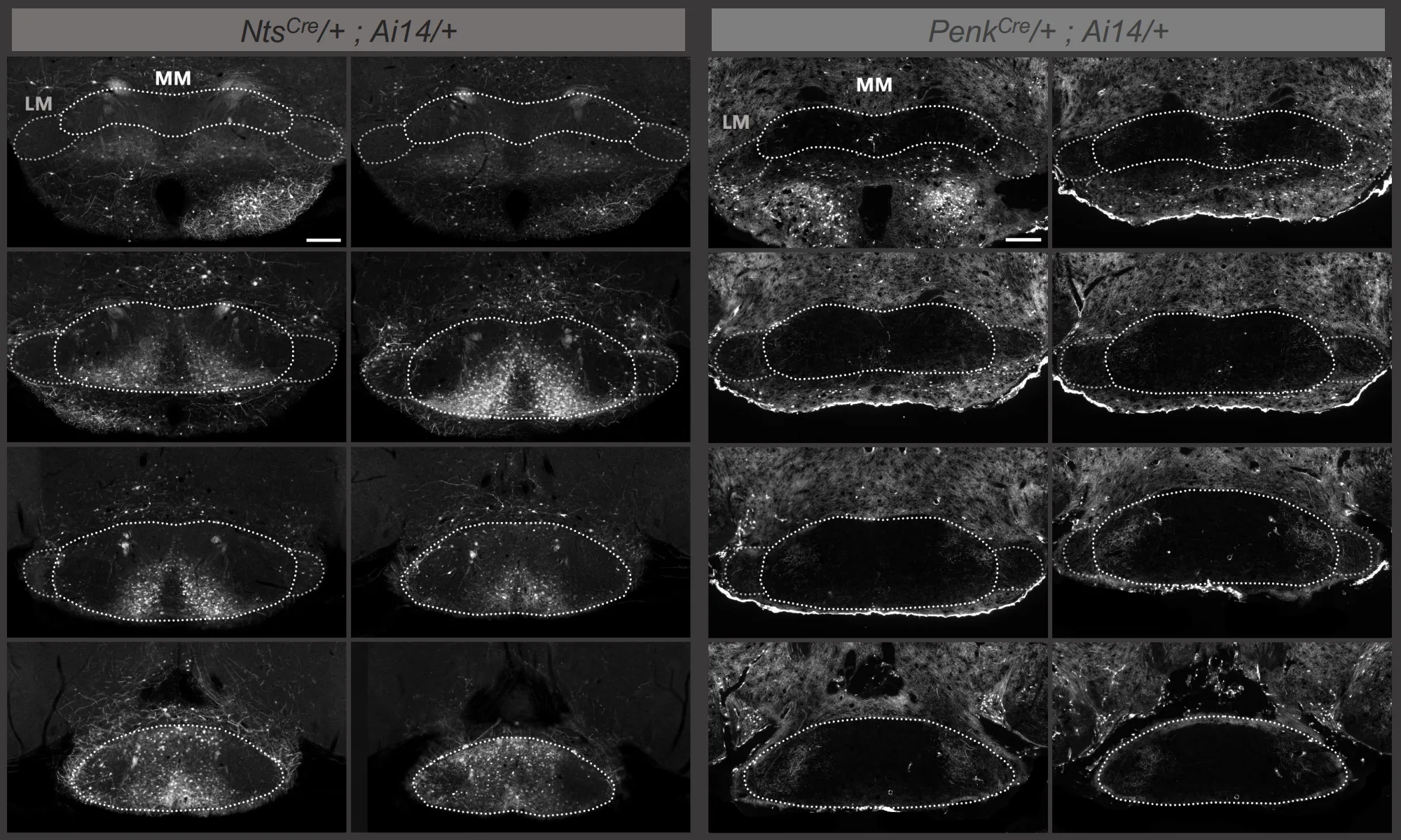
The medial mammillary body (MM) contains molecularly distinct neuronal populations whose anatomical organization and circuit connectivity remain incompletely understood. This project aims to characterize the spatial distribution and circuit organization of Penk⁺ and Nts⁺ neurons in the MM. Using Allen Brain Atlas in situ hybridization data together with Penk-Cre;Ai14 and Nts-Cre;Ai14 reporter mice, we found that Nts⁺ neurons are relatively abundant and concentrated in the middle-to-posterior MM, whereas Penk⁺ neurons are sparse and largely restricted to a narrow region in the anterior MM. We next attempted to identify upstream inputs to Penk⁺ MM neurons using a Cre-dependent monosynaptic rabies tracing strategy, with the interanteromedial thalamic nucleus (IAM) or anteromedial thalamic nucleus (AM) serving as downstream targets. However, reliable starter-cell labeling could not be achieved because of the highly restricted distribution of Penk⁺ neurons, the high precision required for viral targeting, and substantial off-target infection in surrounding Penk-rich regions. These results establish the distinct spatial organization of Penk⁺ and Nts⁺ neuronal populations in the MM and identify key technical constraints for projection-defined circuit tracing in this small hypothalamic structure. Future work will focus on improving viral targeting accuracy and extending circuit mapping to the more abundant Nts⁺ population.